|
Oculoplastic procedures include a wide range of surgical procedures that deal with the eye socket (orbit), eyelids, tear ducts, and the face. They include the repair of orbital fractures, tear duct obstructions, droopy eyelids, removal of tumors in and around the eyes, and facial rejuvenation treatments such as eye lifts, brow lifts, face lifts and laser skin resurfacing.
Oculoplastic procedures often provide both functional and aesthetic results. An example is blepharoplasty through which we remove excessive, droopy eyelid skin to rejuvenate the appearance, often with the added benefit of increased peripheral vision.
Please read further to learn about the oculoplastic procedures we offer.
Skin Cancer/Lesions
Our doctors are highly experienced in the care of skin cancers and lesions. Skin cancers can occur on the eyelids, nose, in the corner of the eye and in the area of the eyebrows. They can also invade the eye socket (orbit). Typically, they appear as nodules (an overgrowth of tissue), often having an ulcerated center with a pearly border. Skin cancer often seems to a patient like they have a sore that won't heal.
Skin cancer treatment is managed in two ways: Excision (surgical removal) of the lesion or irradiation. Cancer must be completely removed or destroyed to eliminate its continued growth or recurrence. Our doctors will generally perform surgical excision but if the tumor is too large in size or if the patient is not a good candidate for surgery, irradiation will be necessary.
If you have a sore that won't seem to heal or believe that you may have a cancerous lesion, please don't delay. Call our office today so that our understanding and gentle doctors can evaluate your needs and help you to receive appropriate treatment. Early diagnosis promotes your best outcome.
 Back to Top Back to Top
BOTOX®
BOTOX (botulinum toxin type A) is an FDA approved form of the botulinum toxin, proven to be safe and effective with no threat of causing the illness, botulism. BOTOX works by blocking nerve impulses to the muscles.
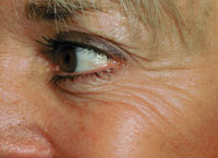
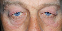
Before
|
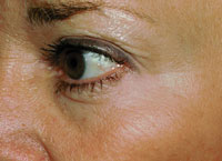
After Botox® Treatment
|
BOTOX is able to be used to treat numerous ophthalmic conditions including:
-
Blepharospasm — In blepharospasm, one or both of the eyelids spasm closed, forcefully and repeatedly, sometimes even resulting in temporary vision loss. Blepharospasm has been associated with fatigue and stress, although its actual cause remains unknown. BOTOX, injected in small doses in the affected upper and lower eyelids, can provide relief of this condition.
Treatment effects generally begin within 1 to 14 days and last for 3 to 6 months. Depending on severity and recurrence, repeat treatments may be necessary every 3 to 6 months.
-
Hemifacial spasm — Hemifacial spasm is a condition in which powerful, painful contractures occur to one half of the face. Neuroimaging, such as CT or MRI, may be recommended since hemifacial spasm may be due to an intracranial tumor or vascular compression of the facial nerve.
Treatment can include surgery; ablation (precise surgical removal) of branches of the facial nerve; or the injection of BOTOX. If the underlying cause cannot be found or is not treatable through other means, BOTOX may successfully treat the symptoms of hemifacial spasm. The doctor uses a tiny needle to administer BOTOX in select locations on the face. Retreatment is generally required every 3 to 6 months.
-
Facial wrinkles — BOTOX is very helpful in the treatment of wrinkles in the forehead and adjacent to the eyes. Using an extremely fine needle, the doctor places minute doses of BOTOX in the needed areas. This treatment provides an excellent alternative to more invasive cosmetic surgery procedures. Effects last approximately 3 to 6 months.
Back to Top
Ectropion
An ectropion is usually associated with aging but additionally occurs as a result of other surgeries or scarring, as secondary to facial nerve paralysis (Bell's palsy), and can also be congenital. Almost always affecting the lower eyelid, ectropion is an outwardly turned (everted) eyelid. When not repaired, it can lead to thickening of the mucosal surface on the inside of the eyelid (the conjunctiva). This can result in inflammation to the eye itself and a danger to its health.
Repair for ectropion is based upon its underlying cause. When associated with aging, known as involutional ectropion, the doctor will generally shorten and tighten the lower eyelid, correcting the outwardly turned lid. This surgery typically consists of an incision at the lateral (outside) corner of the eye. A small segment of skin is excised from the lid and the eyelid is then reconnected to the underlying tissues and the upper eyelid. Only a few sutures are usually required at the outside corner of the eye and are generally removed in 7 to 14 days. The condition almost always resolves immediately. The procedure causes little or no discomfort. Some mild swelling and bruising may follow the procedure and should disappear within approximately two weeks.
Cicatricial ectropion, which is the result of scarring, usually occurs following another surgical procedure of the eyelids or face, particularly excision of skin lesions such as skin cancers. For repair of this type of ectropion, skin grafting is often required. Skin graft donor sites are generally from either the patient's upper eyelid or from behind the ear. These donor sites provide grafts which closely match the skin of the patient's lower eyelid. Within two weeks of surgery, both the surgical site and the donor site will typically heal well.
Back to Top
Entropion
A condition occurring primarily due to aging, an entropion is an inwardly turned (inverted) eyelid. Entropion usually affects the lower rather than the upper eyelids. It can cause irritation by allowing the eyelashes to rub on the ocular surface. Symptoms can include eye or lid pain; redness or pinkness of the eye; the sensation of a foreign body in the eye; tearing; itching; and vision loss. It is the result of an imbalance between the muscle groups which control the affected eyelid.
Repair of age-associated involutional entropion is achieved through a number of different procedures. The doctor will generally use an incision at the lateral (outside) corner of the eye or right under the lower eyelashes, followed by the placement of sutures. The imbalance of muscle groups in the eyelid is rectified through tightening of the tissues. Patients may expect some minor bruising or swelling which should disappear in one to two weeks after surgery. Generally, removal of the sutures will be necessary. In most cases, the problem is resolved upon completion of surgery and there is little or no postoperative discomfort.
Back to Top
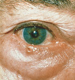
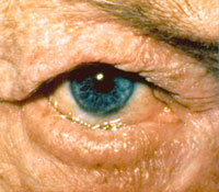
Ptosis
Repair for Droopy Eyelid
Ptosis surgery helps to correct droopy eyelid. In this condition, the upper eyelid droops down; sometimes even over the pupil, blocking vision. Ptosis can occur due to poor function of the levator muscle, located in the upper eyelid, which elevates the lid. It can also develop due to injury, aging, or the aftereffect of other eye surgery. Sometimes ptosis is the result of a neurological disorder, eye tumor or a systemic disease such as diabetes. Infants and children also suffer ptosis. One or both eyes may require treatment.
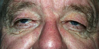
Before: Droopy Eyelids
|

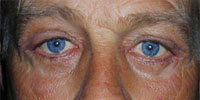
After: Ptosis Surgery
|
In infants and children the surgery is performed under general anesthesia. In adults, it is performed using local anesthesia. During the procedure, the doctor will make an incision in the drooping eyelid and carefully advance and tighten the levator muscle. In awake patients, the eyelid may be compared to the lid height of the opposite eye. With those under general anesthesia, the doctor uses careful measurements and examination to assure the correct height.
Children with poor levator muscle function may require the construction of a frontalis sling. In this more complex procedure, the doctor utilizes muscles of the brow or forehead to assist in the elevation of the upper eyelid. After the correct lid height has been secured, the eyelid incision is closed with tiny sutures.
Postoperative Care
During the 48 hours following surgery, cold packs may need to be applied to the treated eyelid. The doctor may also recommend the application of antibiotic ointment to the incision. Some bruising and swelling may obscure the elevation of the eyelid, but it is often noticeable immediately following surgery. Necessary removal of sutures will occur about one week after surgery. In procedures on children, the doctor will often use absorbable sutures. Any resultant bruising and swelling should disappear in two to three weeks. Some patients may require further adjustment of the sutures for better alignment of lid height. Most patients enjoy a rapid recovery with good results from this procedure.
Back to Top
Probing and Irrigation for Blocked Tear Duct
A relatively common condition in infants, congenital nasolacrimal duct (NLD) obstruction occurs when the nasolacrimal (tear drainage) duct fails to open into the nose. Tears, mucous and bacteria are unable to be cleared from the eye by draining down through the nose as they usually would. This sticky debris then backs up in the eyelid and may be associated with a mild infection along the tear drainage pathway, although rarely jeopardizing the eye itself. In most cases, the obstruction is at the lower end of the duct, right before it enters the nose. Gentle massage of the lacrimal (tear) sac sometimes forces the fluid down the tear duct, clearing the blockage. Topical antibiotic drops may be prescribed for those infants with excessive discharge or crusting around the eye.
There is a high incidence of the tear ducts opening spontaneously by one year of age and parents may elect to wait and see if the condition resolves itself. If immediate intervention is deemed necessary or if obstruction persists, a probing and irrigation procedure may be performed.
The Probing and Irrigation Procedure
If tearing persists, it may be necessary to open the tear duct through a procedure known as probing and irrigation, performed under brief general anesthesia. In this treatment, a thin, blunt metal wire is passed gently through the nasolacrimal drainage system to open the obstruction and then fluid is irrigated through the system. Afterward, some blood-tinged nasal secretion or tears are common, but the infant will experience no pain following the procedure. For up to one week there may be a discharge from the eye and antibiotics may be prescribed. In the few infants where obstruction remains or reoccurs, a repeated probing may be performed. Should the blockage still remain, silicone tubes may be placed in the nasolacrimal duct until the opening is successfully secured, usually within three to six months following placement.
Back to Top
DCR (Dacryocystorhinostomy)
For Blocked Tear Duct
Tears are manufactured in part by the lacrimal gland, a small almond-shaped structure located just above the outer corner of the eye. Each time we blink, tears are drained from the eye into the nose through the tear drainage system.
There are two tiny openings to the tear drainage system, one in the upper eyelid and one in the lower eyelid, called puncta. They are situated in the corner of the eyelid closest to the nose. Tears enter through the puncta into little ducts known as canaliculi and flow into the lacrimal sac on the side of the bridge of the nose. They flow down through the lacrimal sac through the nasolacrimal duct into the nose. Generally, it is toward the bottom of the nasolacrimal duct that obstructions occur.
The DCR Procedure
DCR (dacryocystorhinostomy) is the treatment usually recommended when tearing is diagnosed due to acquired obstruction of the nasolacrimal (tear) duct in adults. During this procedure, the tear drainage pathways are reattached to the inside of the nose to create a new drainage pathway for the tears. Very small plastic stents (tubes) are placed to prevent scarring from blocking the new openings. The stents remain in place for a few months and are then removed in the office with little or no discomfort and no need for anesthesia.
Back to Top
Blepharoplasty
Eyelift
Blepharoplasty is a surgical procedure to correct drooping upper eyelids and puffy bags below the eyes which can cause an aged or tired look. Some patients also suffer diminished peripheral vision because of the overhanging skin. The procedure won't remove "crow's feet", raise sagging eyebrows, or eliminate dark circles beneath the eyes. However, it can enhance the appearance, widen the peripheral field of vision, and relieve eye strain from using forehead muscles to raise the upper eyelids.
Before
|
After Eyelift
|
Blepharoplasty may represent an increased risk to those patients with certain medical conditions such as diabetes, dry eye syndrome, Grave's disease, and high blood pressure. Glaucoma or a history of retinal detachment can also impact this surgery. The doctor will discuss your history with you in detail to allow the best recommendations regarding surgery.
The Blepharoplasty Procedure
The procedure is generally performed under local anesthesia, sometimes accompanied by mild sedation. All four eyelids may be treated at once, or just the upper or lower ones. During treatment, you should not have any pain but may experience occasional mild discomfort. The doctor may decide upon general anesthesia; if so, you will sleep through the procedure. Blepharoplasty usually takes from one to three hours, typically based on whether all four eyelids are operated upon or just the upper or lower ones.
Using a scalpel, the doctor makes incisions along the natural creases of the upper eyelids and just below the eyelashes for the lower eyelids. Excess skin, muscle and fat are removed as necessary to bring about the desired result. The incisions are closed with minute sutures, generally removed in approximately 1 week. When excess skin does not need to be removed when operating on the lower eyelids, the doctor may perform a transconjunctival blepharoplasty. A small incision is made on the inside of the lower eyelid to allow the removal of a fat pocket. This produces the desired result and prevents any visible scar.
Postoperative Care
You will probably be able to leave our office shortly after surgery. Bandages are usually not necessary. You can expect swelling and some bruising and should plan to apply cold packs to your operated eyelids for the 48 hours following surgery. Bruising and swelling generally decrease the first few days after surgery but can remain present for up to one month.
For the first week following surgery, your eyes may feel tight and sore and the doctor may prescribe oral pain medications for the first few days. Sometimes the eyes become dry and a bit irritated. In this case, the doctor will probably recommend the application of non-preserved lubricating tears or lubricating ophthalmic ointment. If swelling is significant, the vision may be blurred or even double; but this will resolve when the swelling subsides.
Makeup can be applied to the eyelids again from 7 to 10 days after surgery.
Results
You will begin to see the results of your surgery after the first couple of weeks, but your best results won't occur for up to 6 months. Your incision lines will fade, becoming nearly invisible.
Back to Top
Ocular Prosthetics
False Eyes
In some cases, traumatic accidents and treatment of ocular and orbital cancers, blind and painful eyes, and other conditions cause the need for reconstruction of the orbit with placement of an orbital implant, or false eye.
Today, with advances in both surgical techniques and aesthetic materials, excellent cosmetic results can be obtained. The muscles and other natural tissues of the eye are preserved to hold prosthetic eye in place. This also encourages natural-looking movement of the eye.
If you should require ocular prosthetics, the doctor will clarify all of the treatment necessary and answer any questions you may have so that you feel confident about your surgery.
Back to Top
Latisse®
For Longer, Thicker Eyelashes
Latisse was launched in 2009 by Allergan, Inc. and is FDA-approved for cosmetic use. Known by the generic name bimatoprost ophthalmic solution, the drug has been used to control internal eye pressure to prevent potential eye damage from glaucoma, but was also noted to result in eyelash growth.
Latisse works gradually to encourage the growth and thickness of eyelashes- with full results after 12-16 weeks. Once you have begun treatment, you simply continue to apply the topical solution each night according to directions to maintain results. Latisse works from the inside out and as you continue treatment, you'll start to see changes in the length , thickness and darkness of your lashes. After week 16, you'll see the full effect of Latisse- and so will your family and friends!
Please speak with us about this exciting new treatment. We will be happy to answer any questions you may have and schedule an appointment to get you started on your way to longer, fuller more beautiful lashes.
Back to Top
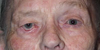
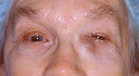
Trichiasis®
Trichiasis is a condition where the eyelashes are misdirected and grow inwards toward the eye. This can occur secondary to an abnormal eyelid margin or with a normal eyelid margin. The inward turning of the lashes cause irritation and damage to the cornea and conjunctiva.
Causes
-
Chronic blepharitis
-
Epiblepharon
-
Trachoma
-
Rare skin disorders of skin and mucous membranes
-
Abnormal eyelid margin
-
Previous eyelid surgery
-
Trauma
-
Idiopathic
Symptoms
-
Ocular irritation
-
Redness
-
Tearing
Treatment
If the trichiasis is limited to a single/ few lashes they can be removed with tweezers in office, with the possibility of recurrence. If there are multiple lashes and/ or recurrent lashes, you can have permanent removal with a laser procedure or electrolysis.
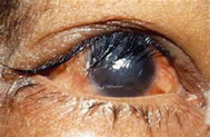
Trichiasis
|
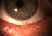
Trichiasis
|
Back to Top
|