|
The macula is a small, extremely important area in the back of the eye. It provides the sharp, central vision necessary to see in fine detail, to read, and to drive. Macular degeneration, also know as age-related macular degeneration (ARMD) involves the breakdown of the macula.
In Caucasians over the age of 65, macular degeneration is the primary cause of severe vision loss. It develops in many older people as part of the natural aging process of the body. It isn't known why it develops and no treatment has yet been found to be uniformly effective.
Macular degeneration can cause a sudden and severe loss of vision in the middle of the visual field. It is important to have regular eye exams by an ophthalmologist, particularly after age 65. With proper examination, our doctors are able to detect macular degeneration in its early stages.
Symptoms
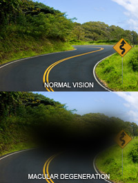
Macular degeneration reduces vision in the central part of the retina but generally does not affect peripheral (side) vision. If straight lines appear crooked, an empty or dark area appears in the center of your vision, or words look blurry as you read, you may be experiencing symptoms of macular degeneration. It is possible to lose vision in one eye while continuing to see well out of the other. If both eyes have macular degeneration, central vision problems will be apparent more quickly.
 Back to Top Back to Top
Testing
Testing for ARMD includes:
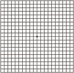
Amsler Grid Testing — This simple screening test is used to assess the macula (the center of the retina). The Amsler Grid consists of evenly spaced horizontal and vertical lines printed on black or white paper with a small dot located in the center of the grid for fixation. While staring at the dot, the patient looks for wavy lines and missing areas of the grid. If the lines of grid do not appear straight and parallel or there are missing areas, the doctor will examine the back of the eye (macula) very closely. This test is frequently given to patients for home use to monitor ARMD.
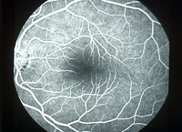
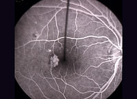
Fluorescein Angiogram — This test enables us to photograph the blood vessels in the back of the eye as a fluorescent dye is injected through the arm or hand into the bloodstream. It is performed to allow the doctor to confirm diagnosis, develop guidelines for treatment, and to maintain a permanent record of the vessels at the back of your eye. The test is especially helpful for management of macular degeneration and diabetic retinopathy.
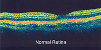
OCT (Ocular Coherence Tomography) — Progressive diseases such as ARMD necessitate the consistent, accurate and reliable detection of change as time goes by. 3-D images of the retina, nerve fiber layer and optic disc, used to diagnose macular edema and glaucoma, are provided by scanning laser technology.
Ophthalmoscopy — An ophthalmoscope, a device with a small light at its end, is used to examine the internal structures of your eye. Used in a darkened room, it allows the doctor to examine the inside of the eye, particularly the optic nerve.
Fundus Autofluorescence Photography — This imaging may allow for early identification in retinal diseases which may not be evident on examination. This is also very helpful to investigate those patients with unknown vision loss or those with family history of hereditary retinal diseases.
Back to Top
Types of ARMD
The two most common types of ARMD are:
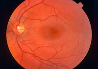
Dry Macular Degeneration (Atrophic)
Caused by aging and thinning of the tissues of the macula, most individuals have the "dry" form of ARMD. In this form, vision loss is usually gradual.
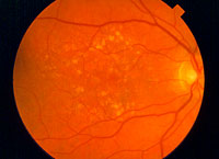
Dry Macular Degeneration
|
Fluorescein Angiogram
|
Treatment
While there is no specific treatment for dry macular degeneration at this time, strong evidence exists that certain vitamins such as those found in the AREDS formula may help prevent or slow its progression. Sunglasses with UV protection may also help to guard against the damaging rays of the sun. Some cases of dry macular degeneration advance to the wet form and you will also be regularly examined to monitor your status. Our doctors will advise you on the best program to follow for your particular case.
Back to Top
Wet Macular Degeneration (Exudative)
Approximately 10% of cases represent the "wet" form of ARMD. It occurs as a result of the formation of abnormal blood vessels at the back of the eye underneath the retina. Those blood vessels leak blood or fluid which blurs central vision. In this form, vision loss can be severe and rapid.
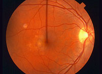
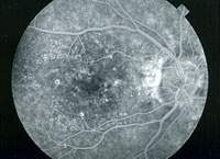
Wet Macular Degeneration
|
Fluorescein Angiogram
|
A common feature of macular degeneration is deposits under the retina called drusen. Vision loss is not generally caused by drusen alone, but when their number or size increase it is usually indication of an increased risk of developing advanced ARMD. Those at risk for developing advanced ARMD have prominent dry ARMD; significant drusen; or, in the wet form, abnormal blood vessels under the macula.
Treatment
While macular degeneration can't be reversed, its impact can be reduced through appropriate management.
Antioxidant vitamins, zinc and copper have been shown to ease its impact in some patients. Laser surgery can be used to treat certain types of wet macular degeneration. This is performed as a brief outpatient procedure in which a focused beam of light is used to slow or stop leaking blood vessels which damage the macula. A combination of a specific drug and laser treatment to slow or stop leaking blood vessels is used in a procedure called photodynamic therapy (PDT).
Another treatment targets a chemical in the body called vascular endothelial growth factor (VEGF) which causes abnormal blood vessels to grow under the retina. Anti-VEGF medications block the VEGF, thereby reducing abnormal blood vessel growth and slowing leakage. Medication is delivered by intraocular injection. This breakthrough treatment is the first to not only halt further damage, but, in some cases, to actually improve the patient's vision. We utilize Lucentis®, Avastin®, and Eylea anti-VEGF medications. Dr. Klug will decide which treatment is right for you.
In spite of advanced medical treatment, the majority of people with macular degeneration experience some vision loss. However, although they cannot restore vision back to normal, these procedures do allow us to help preserve sight for many patients.
Back to Top
|