|
Glaucoma is a leading cause of blindness in the United States, largely because there are no symptoms until its later stages when vision loss is irreversible. The disease damages the optic nerve which is the part of the eye that carries the images you see to the brain. Glaucoma can be well managed through early diagnosis and treatment. It's important to get regular eye exams by an ophthalmologist to maintain the health of your eyes.
Glaucoma Risk Factors
Primary risk factors for glaucoma include:
-
age (60 and older)
-
elevated eye pressure
-
family history of glaucoma
-
African or Hispanic ancestry
-
farsightedness or nearsightedness
-
previous eye injuries
-
steroid use
-
other health problems such as diabetes or migraine headache
Types of Glaucoma
Open-Angle Glaucoma
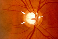
Much like an electric cable containing many wires, the optic nerve consists of nerve fibers which transmit images to the brain. Blind spots develop when glaucoma causes damage to the optic nerve fibers. If the whole nerve is destroyed, blindness occurs.
The most common form of glaucoma in the United States is chronic open-angle glaucoma. Generally, in its early stages, open-angle glaucoma has no symptoms and vision is normal.
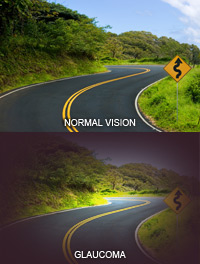
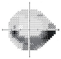
Blank spots start to appear in the field of vision as damage to the optic nerve increases. These blank spots are not noticeable in your daily activities until the optic nerve is seriously damaged and they become large. Blindness results if all of the optic nerve fibers die.
 Back to Top Back to Top
Closed-Angle Glaucoma
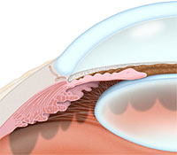
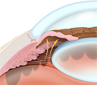
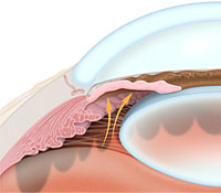
From birth, in some people, the iris (the colored part of the eye) is too close to the drainage angle. Their eyes are often small and farsighted and the iris can be pushed into the drainage angle, completely blocking it. Because fluid cannot exit the eye, pressure builds quickly inside the eye, resulting in an acute, sudden closed-angle attack.
Two-thirds of patients with closed-angle glaucoma develop it over the course of time with no symptoms to warn that an acute attack may occur.
Some symptoms of closed-angle glaucoma are:
-
blurry vision
-
severe eye pain
-
headache
-
rainbow-colored halos around lights
-
nausea and vomiting
If you experience these symptoms, it is a true eye emergency. Please call our office immediately. Without quick treatment, blindness can result.
Back to Top
Other Forms of Glaucoma
Narrowed side vision and optic nerve damage occur in normal-tension or low- tension glaucoma. Diagnosis is more difficult because intraocular pressure tests within normal limits.
Congenital glaucoma causes children to be born with a defect in the angle of the eye which impedes normal fluid drainage. Symptoms in these children are generally obvious, including cloudy eyes, excessive tearing and a sensitivity to light.
Back to Top
Testing and Diagnosis
The best way to detect glaucoma and maintain the overall health of your eyes is through regular eye exams at our office. Testing for glaucoma includes:
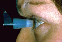
Tonometry Test — The pressure of the eye is measured by the tonometry test. Drops are used to numb the eye and a special piece of equipment measures the pressure.
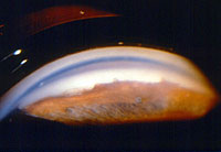
Ophthalmoscopy — An ophthalmoscope, a device with a small light at its end, is used to examine the internal structures of your eye. Used in a darkened room, it allows the doctor to examine the inside of the eye, particularly the optic nerve.
Perimetry Test — This is also known as a visual field test (Humphrey Visual Field). During the test, you will look straight ahead and indicate when a moving light passes your peripheral (side) vision. This test help to draw a map of your vision.
Gonioscopy — A painless eye test, gonioscopy determines whether the angle where the iris meets the cornea is open or closed. This indicates whether either open-angle or closed-angle glaucoma is present.
Pachymetry — Corneal thickness is precisely measured by a non contact method, using optical low coherence reflectometry.
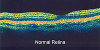
OCT (Ocular Coherence Tomography) — Progressive diseases such as glaucoma necessitate the consistent, accurate and reliable detection of change as time goes by. 3-D images of the retina, nerve fiber layer and optic disc, used to diagnose macular edema and glaucoma, are provided by scanning laser technology.
Optic Nerve/Fundus Photography — Optic nerve photography remains the standard of analysis for imaging of the optic nerve head in patients with glaucoma and those being evaluated for possible glaucoma. The optic nerve head and fundus (the back part of the eye's interior, including the retina) are photographed during successive visits. The resulting images are compared for structural changes which are indicative of the progression of glaucoma.
Not every test is needed for every patient. The tests the doctor determines are necessary for diagnosis may also be repeated on a regular schedule to help track changes in your condition.
Back to Top
Treatment
Vision loss can usually be prevented through treatment, however, the damage caused by glaucoma is irreversible.
Further damage can be prevented through the use of eyedrops, laser surgery, and conventional surgery. Oral medications may be prescribed in some cases.
Eyedrops used daily are generally prescribed to control glaucoma. These drops lower eye pressure by either improving flow through the drainage angle or by decreasing the amount of fluid produced within the eye. Our doctors will discuss the use of these medications and any side effects they may produce.
Regular examinations are very important in the management of glaucoma. Changes to your treatment may be needed from time to time since glaucoma can progress without symptoms.
Back to Top
Glaucoma Procedures
Some of the procedures used to treat glaucoma include:
Surgical Treatment
-
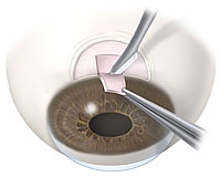
Trabeculectomy (Glaucoma Filtration Procedure) — Used to create a new passageway by which the aqueous fluid within the eye can escape, thereby lowering pressure inside the eye.
-
Glaucoma Drainage Device (Tube Shunt) — These devices are implanted for those patients at high risk for failure with a trabeculectomy procedure, such as those with neovascular glaucoma, glaucoma associated with uveitis, or those under 30 years of age with glaucoma. The shunts are designed to maintain an artificial drainage pathway.
-
iStent Trabecular Micro-Bypass Stent — The iStent is a device that is implanted into the eye during cataract surgery in those who have been diagnosed with mild to moderate glaucoma. This procedure is performed to increase the outflow of the fluid in the eye, decreasing the pressure.
Laser Treatment
-
Laser Peripheral Iridotomy (PI) — Performed almost exclusively for patients with narrow angles, narrow angle glaucoma, or acute angle closure glaucoma, PI involves creating a minute opening in the peripheral iris directly to the anterior chamber of the eye. A successful result corrects the forward-bowing of the iris, opening the angle of the eye to allow aqueous flow and relieve internal pressure.
-
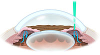
Argon Laser Trabeculoplasty (ALT) — In this procedure, the doctor directs a laser beam into the trabecular meshwork, the primary aqueous drainage region of the eye. This serves to increase drainage of the aqueous fluid out of the eye, lowering the intraocular pressure.
-
Selective Laser Trabeculoplasty (SLT) — This treatment represents a noteworthy advance in the surgical treatment of intraocular pressure (IOP) in patients with open-angle glaucoma. Unlike its precursor, argon laser trebeculoplasty (ALT), SLT is able to be repeated several times. ALT patients can receive two treatments within their lifetime. SLT patients are able to receive two treatments per year. This is because the laser beam circumvents surrounding tissue, leaving it completely undamaged.
-
Endoscopic Cyclophotocoagulation (ECP) — ECP is performed on an out-patient basis. In this procedure, the ciliary body of the eye (located just behind the iris) which produces aqueous fluid is treated with the laser to decrease production of this fluid. This reduces pressure within the eye. This procedure is used in combination with cataract surgery or in those who have already had cataract surgery.
Back to Top
|